Carpal tunnel x ray positioning – Carpal tunnel x-ray positioning plays a pivotal role in accurately diagnosing carpal tunnel syndrome, a common condition that causes pain, numbness, and tingling in the hand and wrist. This comprehensive guide delves into the intricacies of proper positioning techniques, ensuring optimal visualization of the carpal tunnel and aiding in the detection of potential abnormalities.
The Artikel encompasses essential aspects of carpal tunnel x-ray positioning, including hand and wrist positioning, projection selection, radiographic techniques, interpretation, special considerations, and quality control measures. By adhering to these guidelines, healthcare professionals can obtain high-quality images that facilitate accurate diagnosis and effective treatment planning.
Positioning the Hand and Wrist

Proper positioning of the hand and wrist is crucial for an optimal carpal tunnel x-ray. By aligning the hand and wrist correctly, we ensure clear visualization of the carpal tunnel, allowing for accurate diagnosis and treatment.
Hand and Wrist Alignment
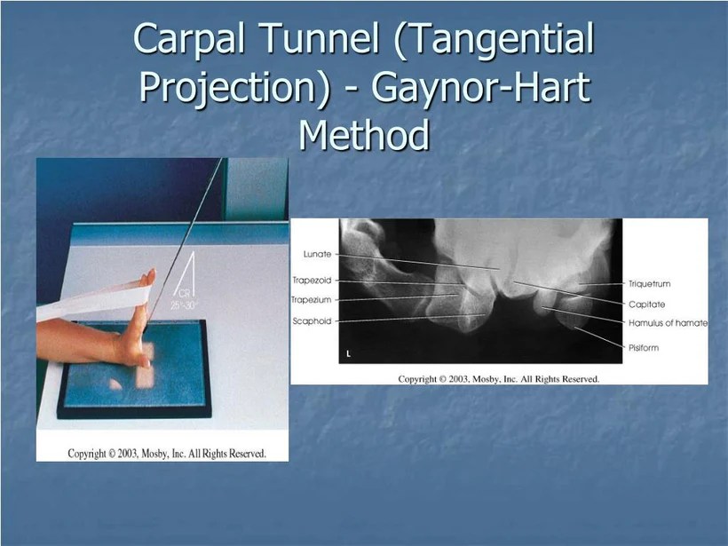
To achieve proper alignment, follow these steps:
- Position the patient’s hand supinated (palm facing up) on the x-ray cassette.
- Flex the wrist at a 90-degree angle to the forearm.
- Align the long axis of the third metacarpal (middle finger) parallel to the x-ray beam.
- Position the ulnar styloid process (the bony prominence on the inner side of the wrist) at the center of the x-ray cassette.
Determining the Optimal Projection

The optimal projection for carpal tunnel x-rays depends on the specific anatomical structures of interest. Different projections provide different views of the carpal tunnel, each with its advantages and disadvantages.
Anteroposterior (AP) Projection
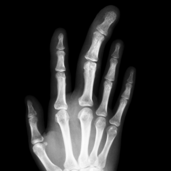
The AP projection is taken with the hand positioned palm down on the x-ray table. This projection provides a clear view of the carpal bones and the distal radius and ulna. It is useful for evaluating the overall alignment of the carpal bones and for detecting fractures or dislocations.
Lateral Projection
The lateral projection is taken with the hand positioned sideways on the x-ray table. This projection provides a view of the carpal tunnel from the side, allowing for visualization of the flexor tendons and the median nerve. It is useful for evaluating the size and shape of the carpal tunnel and for detecting any narrowing or compression.
Carpal tunnel x-ray positioning is a crucial aspect of accurately diagnosing this condition. However, have you ever wondered about the legality of “hello mood” in Alabama? Check out this informative article: is hello mood legal in alabama for more insights.
Returning to our topic, proper positioning of the hand and wrist during carpal tunnel x-ray is essential for optimal image quality and accurate interpretation.
Oblique Projections
Oblique projections are taken with the hand positioned at an angle between the AP and lateral projections. These projections provide views of the carpal tunnel from different angles, allowing for a more comprehensive evaluation of the anatomy. They are useful for evaluating the relationship between the carpal bones and the flexor tendons and for detecting any abnormalities in the soft tissues.
Choosing the Most Appropriate Projection, Carpal tunnel x ray positioning
The most appropriate projection for capturing the carpal tunnel anatomy depends on the specific clinical question being asked. In general, the AP projection is used for a general overview of the carpal tunnel, while the lateral and oblique projections are used for more detailed evaluation of specific structures.
Using Proper Radiographic Techniques

To obtain high-quality carpal tunnel X-rays, proper radiographic techniques must be employed. These include selecting the appropriate tube-to-image distance, kilovoltage (kVp), milliamperage (mA), and exposure time.
Tube-to-Image Distance
The tube-to-image distance should be set to approximately 40 inches (100 cm) to minimize geometric distortion and ensure sharp images.
Kilovoltage (kVp)
The kVp should be set between 50 and 60 kVp to provide sufficient penetration through the wrist bones while minimizing radiation exposure.
Milliamperage (mA)
The mA should be adjusted to achieve an optimal density on the X-ray image. Typically, a range of 10 to 20 mA is used.
Exposure Time
The exposure time should be set to ensure adequate penetration and avoid motion blur. This will vary depending on the kVp and mA settings used.
Interpreting Carpal Tunnel X-Rays: Carpal Tunnel X Ray Positioning
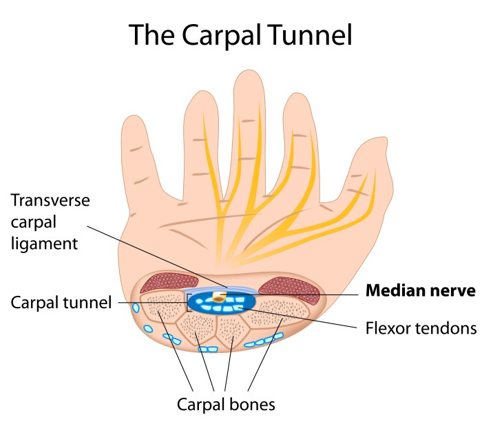
Interpreting carpal tunnel x-rays involves examining the images to identify normal anatomy and detect signs of carpal tunnel syndrome (CTS). CTS is a condition characterized by compression of the median nerve as it passes through the carpal tunnel in the wrist.
Identifying Normal Anatomy
- Bones:The x-ray should show the bones of the wrist and hand, including the carpal bones, metacarpals, and phalanges.
- Carpal Tunnel:The carpal tunnel is a narrow space formed by the carpal bones and the transverse carpal ligament. It should be clearly visible on the x-ray.
- Median Nerve:The median nerve is not directly visible on x-rays. However, its position can be inferred from the location of the carpal tunnel.
Detecting Signs of Carpal Tunnel Syndrome
- Narrowing of the Carpal Tunnel:CTS can cause the carpal tunnel to become narrower, which can be seen on x-rays as a decrease in the space between the carpal bones and the transverse carpal ligament.
- Soft Tissue Swelling:Inflammation associated with CTS can cause soft tissue swelling in the carpal tunnel, which can be seen on x-rays as an increase in the density of the soft tissues.
- Osteophytes:Bone spurs (osteophytes) can form on the carpal bones in some cases of CTS, which can be seen on x-rays as small, pointed projections of bone.
Ruling Out Other Conditions
Other conditions can mimic the symptoms of CTS, such as:
- Tendonitis:Inflammation of the tendons in the wrist can cause pain and discomfort similar to CTS.
- Arthritis:Degenerative changes in the joints of the wrist can also cause pain and stiffness that can resemble CTS.
- Fractures:A fracture of the wrist or hand can cause symptoms that overlap with CTS.
X-rays can help rule out these other conditions by providing a clear view of the bones and soft tissues of the wrist and hand.
Special Considerations

In specific patient populations, additional considerations are necessary for obtaining optimal carpal tunnel x-rays:
Patients with Deformities or Previous Injuries
Deformities or previous injuries can alter the anatomy of the hand and wrist, affecting the positioning and interpretation of the x-rays.
- Proper positioning may require special maneuvers or modifications to accommodate the deformity or injury.
- Interpretation should take into account the altered anatomy and potential for superimposed structures.
Patients with Obesity
Obesity can present challenges in obtaining clear x-rays due to increased soft tissue.
- Increased exposure may be necessary to penetrate the thicker tissue.
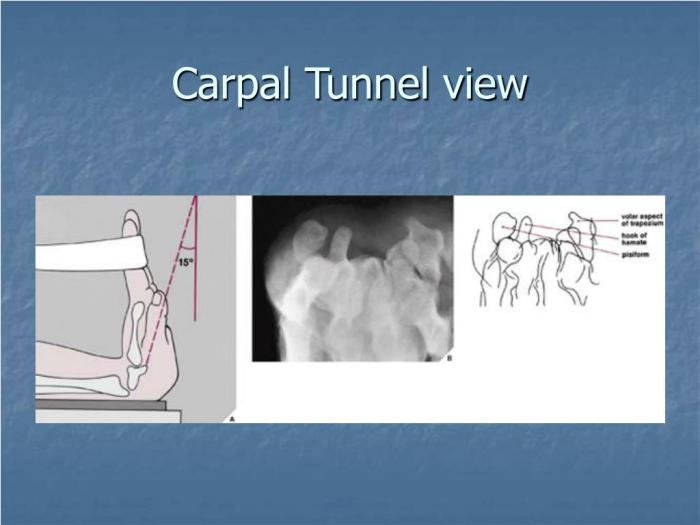
- Alternative positioning techniques, such as the “oblique” or “skyline” view, may be employed to minimize soft tissue overlap.
Patients with Motion Limitations
Motion limitations can restrict the ability to position the hand and wrist optimally.
- Alternative positioning techniques or assistive devices may be necessary to achieve the desired projection.
- Collaboration with the patient and careful manipulation may be required to obtain the best possible images.
Quality Control and Patient Safety

Ensuring the quality of carpal tunnel x-rays is crucial for accurate diagnosis and effective patient care. Quality control measures help maintain the reliability and consistency of the images, minimizing the risk of misinterpretation and incorrect treatment decisions.
Patient safety is also paramount. Radiation exposure during x-ray imaging should be kept to a minimum, and proper positioning techniques must be employed to avoid discomfort or injury to the patient.
Quality Control Measures
- Regular calibration and maintenance of x-ray equipment
- Use of standardized imaging protocols and techniques
- Quality assurance programs to monitor and improve image quality
Patient Safety Considerations
- Minimizing radiation exposure through appropriate shielding and optimization of exposure parameters
- Proper positioning techniques to avoid unnecessary discomfort or injury
- Clear communication with the patient to address any concerns or questions
Commonly Asked Questions
What is the optimal projection for visualizing the carpal tunnel?
The anteroposterior projection is generally preferred for visualizing the carpal tunnel, as it provides a clear view of the carpal bones and soft tissues.
How do you ensure proper hand and wrist positioning during the examination?
The hand should be placed in a neutral position with the fingers extended and the wrist slightly flexed. The wrist should be supported by a firm surface to prevent movement during the exposure.
What are some special considerations for carpal tunnel x-rays in patients with obesity?
In obese patients, additional projections may be necessary to obtain adequate visualization of the carpal tunnel. A lateral projection can be helpful in these cases.